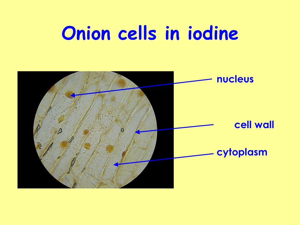
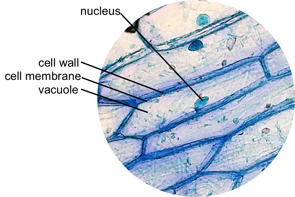
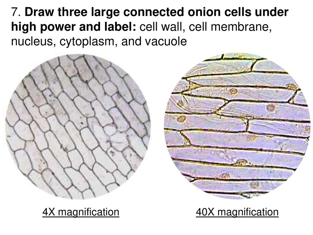
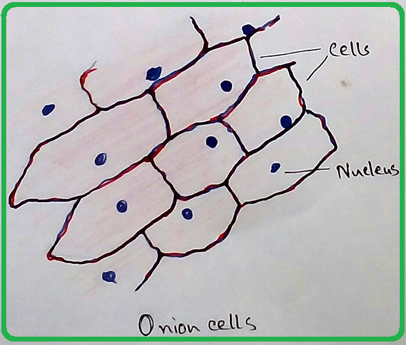
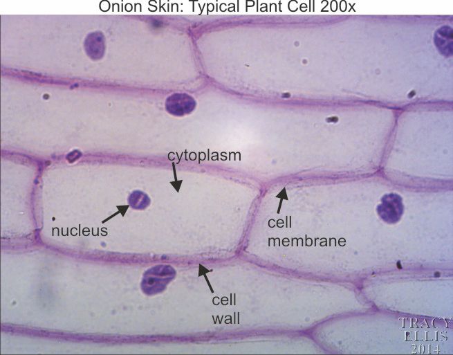
40 onion cells under microscope with labels
ONION CELLS VIDEO - YouTube Sep 26, 2013 ... Video shows how to make a wet mount slide to view onion cells under the microscope. School Science/How to prepare an onion cell slide - Wikibooks Tissue from an onion is a good first exercise in using the microscope and viewing plant cells. The cells are easily visible under a microscope and the ...
VIEWING PLANT CELLS UNDER THE MICROSCOPE: onion cell ... Set aside a clean microscope slide. 2. Carefully cut away a small, single layered piece of onion (1-2 cm wide). 3. Peel the thin layer of skin.

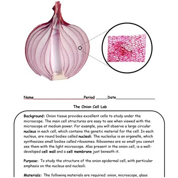
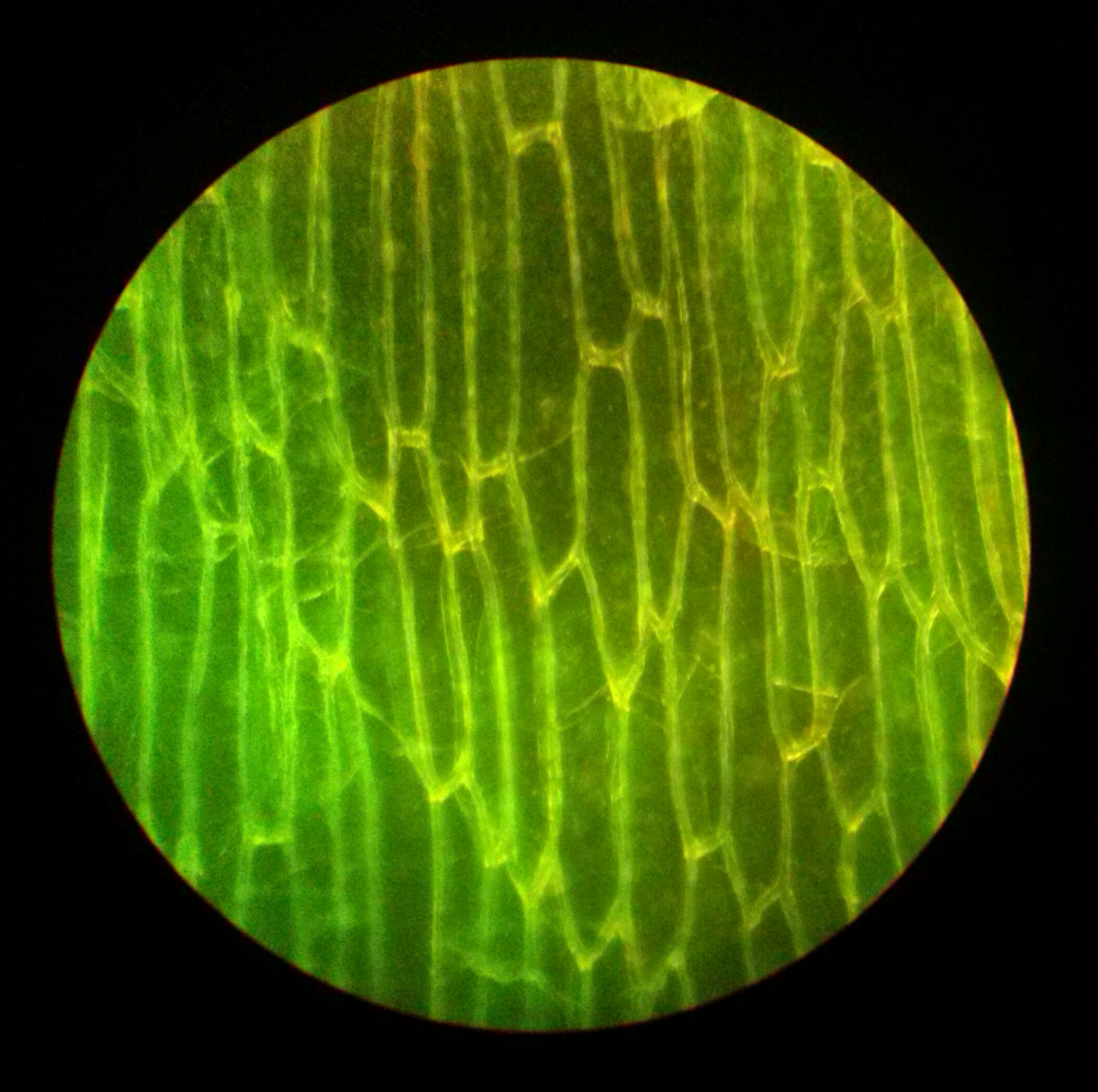
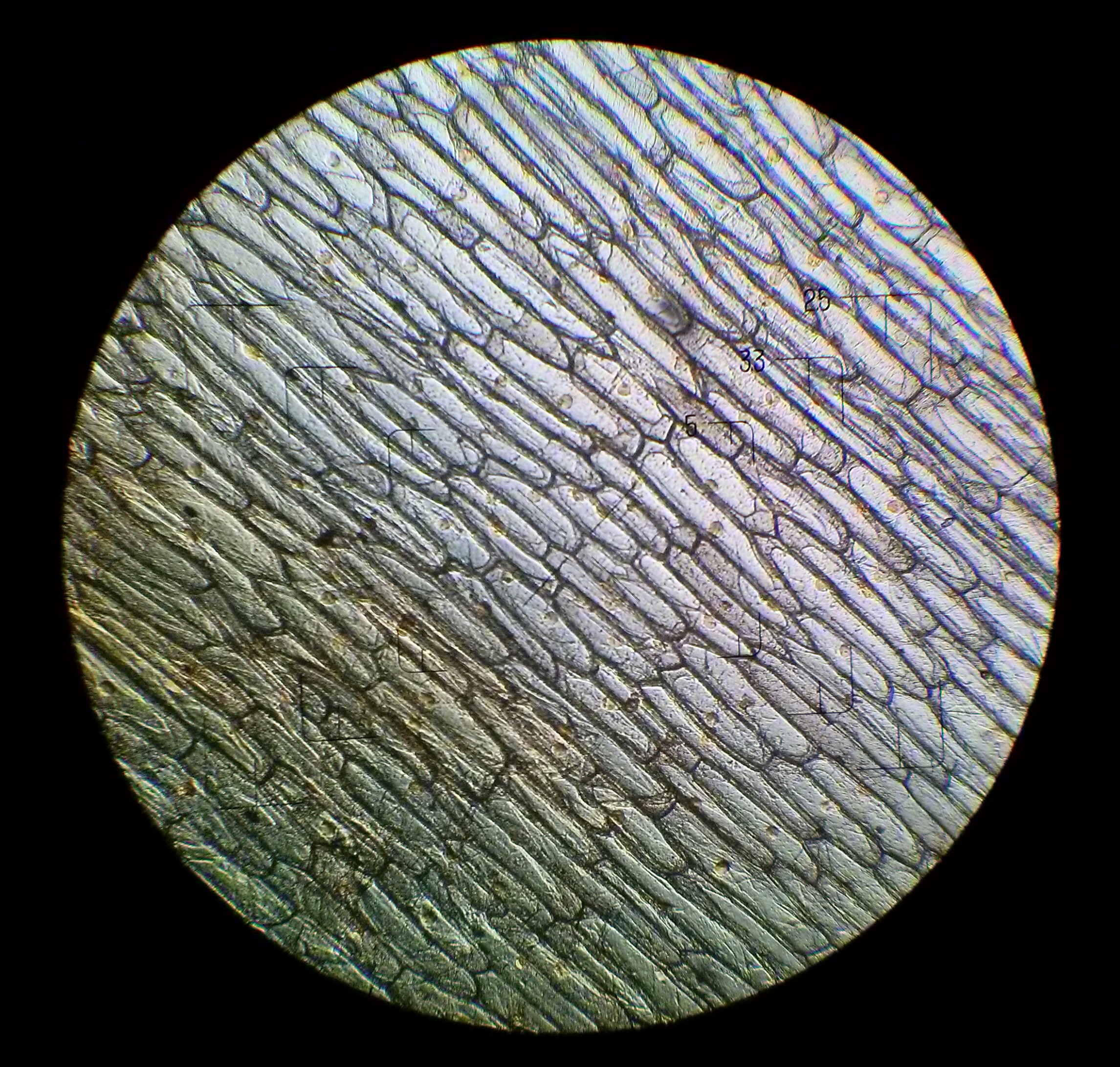
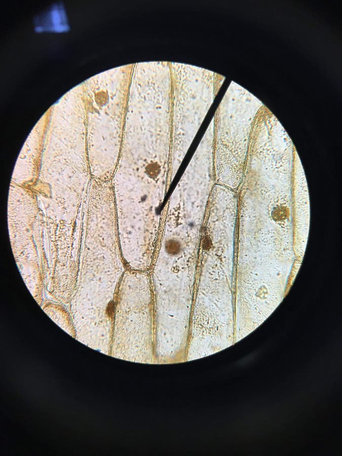
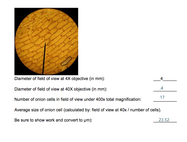
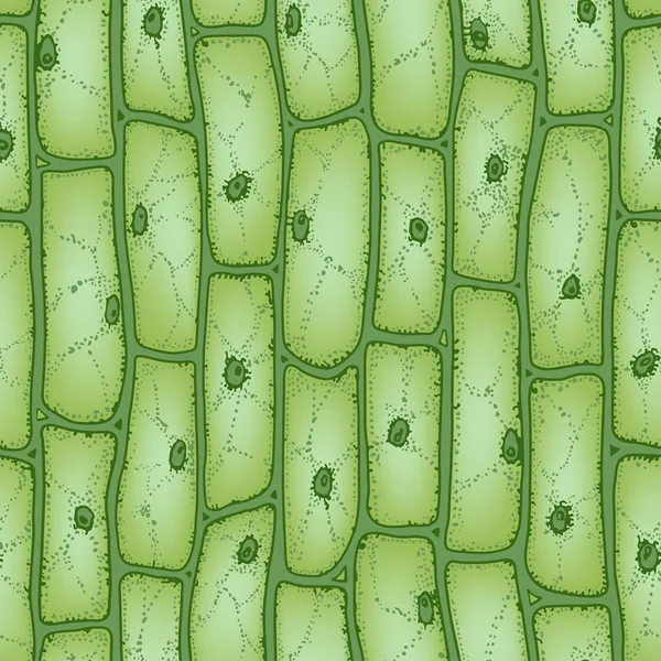
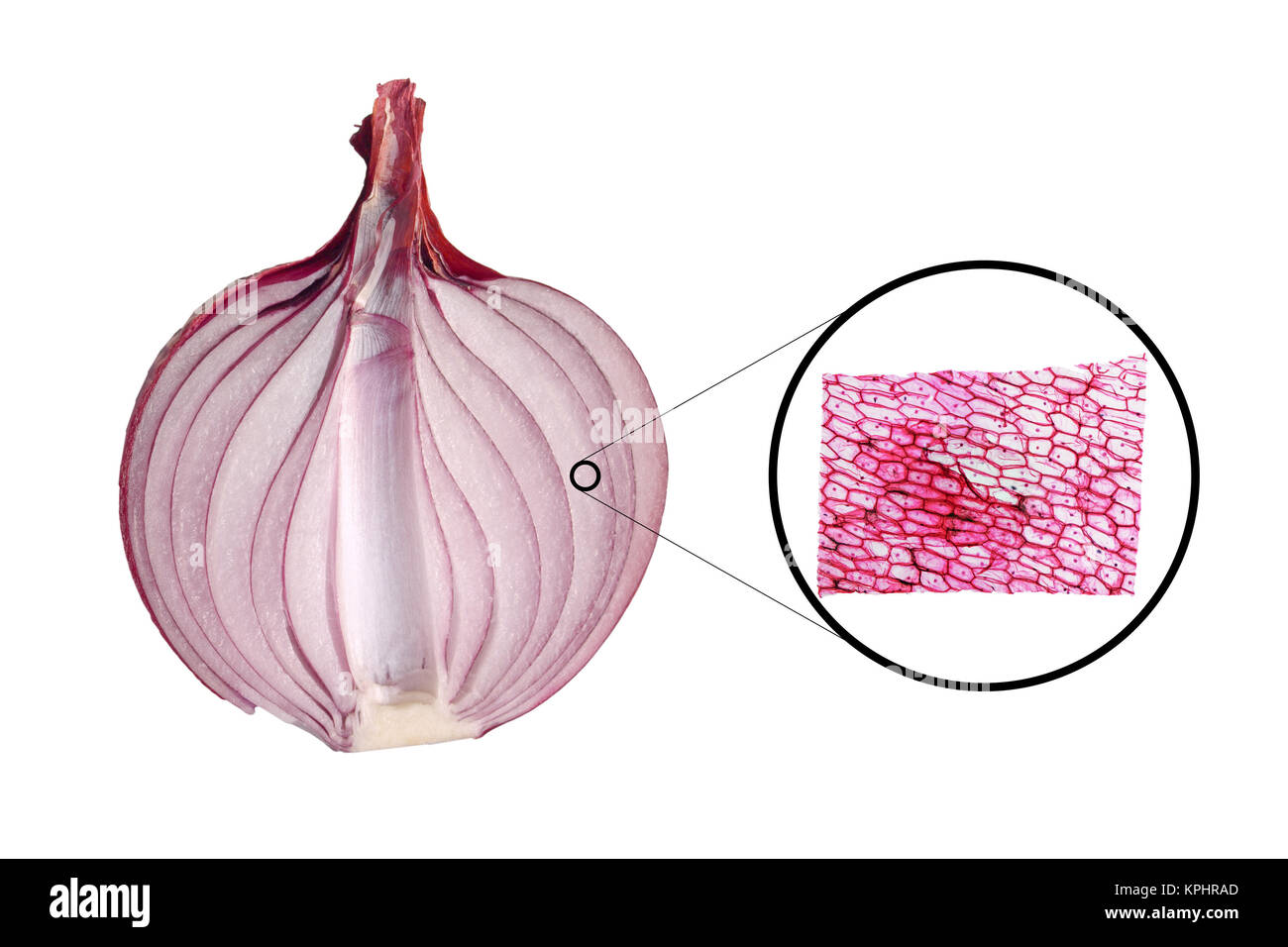
Onion cells under microscope with labels
Onion Cells Under a Microscope - Requirements/Preparation ... An onion is made up of layers that are separated by a thin membrane. For this experiment, the thin membrane will be used to observe the onion cells. It can ... Epidermal onion cells under a microscope. Plant cells ... - Pinterest Feb 3, 2017 - The Biology Lab Primer is an innovative approach to teaching biology concepts in the lab. The Biology Lab Primer reiterates core information ... How to Observe Onion Cells under a Microscope - Blog, She Wrote Dec 19, 2015 ... Make a wet mount slide. Observe an onion cell under the microscope. Record our observations. Materials Needed for Observing Onion Cells. You ...
Onion cells under microscope with labels. allinonehomeschool.com › science-year-1Science — Biology – Easy Peasy All-in-One Homeschool See if you can see the parts of a bulb in an onion or garlic clove. Finish reading the rest of the case. Keep clicking next. Level 5-8. Read “What is a bulb?” and follow the directions. Cut open an onion or garlic clove. These are bulbs. Plants can be grown from these. Do you ever see green leaves coming out of your onions or garlic? › Tags › SatelliteSatellite News and latest stories | The Jerusalem Post Mar 08, 2022 · The Jerusalem Post Customer Service Center can be contacted with any questions or requests: Telephone: *2421 * Extension 4 Jerusalem Post or 03-7619056 Fax: 03-5613699 E-mail: [email protected ... Onion Epidermis Onion epidermis, at 100X, iodine stain. Onion epidermal cells, iodine stain, 400X. The nucleus of an onion epidermal cell, 1000X magnification. en.wikipedia.org › wiki › Scanning_electron_microscopeScanning electron microscope - Wikipedia History. An account of the early history of scanning electron microscopy has been presented by McMullan. Although Max Knoll produced a photo with a 50 mm object-field-width showing channeling contrast by the use of an electron beam scanner, it was Manfred von Ardenne who in 1937 invented a microscope with high resolution by scanning a very small raster with a demagnified and finely focused ...
sciencequiz.net › newjcscience › jcbiologyThe Cell - ScienceQuiz.net The diagram shows a group of onion cells. The parts labelled A, B and C respectively are ... The diagram shows a plant cell as seen under a microscope. Two of the ... Onion Cell Microscope Slide Experiment - YouTube Dec 5, 2015 ... Try not to cry while doing this experiment! Learn how to prepare an onion cell microscope slide!--Sci Files posts all kinds of science ... › pmc › articlesPlant Products as Antimicrobial Agents - PMC Determination of viral infectivity under two conditions: I. In cultured cells during virus multiplication in the presence of a single compound (A-S) or a mixture of compounds, e.g., plant extracts (A-M). II. After extracellular incubation with a single compound (V-S) or a mixture of compounds (V-M). › environmentEnvironment - The Telegraph Oct 19, 2022 · Water meters should be compulsory and bills should rise, says new Environment Agency chairman. Alan Lovell says households consume too much water and metering is needed to encourage them to cut ...
Preparation and scientific drawing of a slide of onion cells including ... exercise you will make a wet mount on a microscope slide, look at the cells using each objective lens and identify the features of the cell visible under ... issuu.com › cupeducation › docsCambridge Lower Secondary Science Learner's Book 7 sample - Issuu Oct 13, 2020 · Try not to get air bubbles under the cover slip. 6 Turn the objective lenses on the microscope until the smallest one is over the hole in the stage. ... • I saw onion cells down the microscope ... 1,947 Onion Cell Images, Stock Photos & Vectors | Shutterstock Onion epidermis with large cells under light microscope. Clear epidermal cells of an onion,. Onion epidermis - microscopic view Stock Photo. How to Observe Onion Cells under a Microscope - Blog, She Wrote Dec 19, 2015 ... Make a wet mount slide. Observe an onion cell under the microscope. Record our observations. Materials Needed for Observing Onion Cells. You ...
Epidermal onion cells under a microscope. Plant cells ... - Pinterest Feb 3, 2017 - The Biology Lab Primer is an innovative approach to teaching biology concepts in the lab. The Biology Lab Primer reiterates core information ...
Onion Cells Under a Microscope - Requirements/Preparation ... An onion is made up of layers that are separated by a thin membrane. For this experiment, the thin membrane will be used to observe the onion cells. It can ...













Post a Comment for "40 onion cells under microscope with labels"