41 adipose tissue with labels
Brown Adipocytes - Yale University Brown adipose tissue is typically found in large amounts in newborns and some hibernating animals and is important as a source of energy. The cells in brown fat contain numerous and very distinct lipid droplets. The presence of an uncoupling protein in these cells causes the generation of heat that allows for non-shivering thermogenesis. Lab Notes Home Page - Kentucky Community and Technical ... Types of Connective Tissue. A). Connective Tissue Proper. 1). Loose Connective Tissue. i). Areolar Connective Tissue. Matrix with fibroblasts supported by gel membrane with collagenous & elastic fibers. ii). Adipose Tissue (fat) Tightly packed & highly vascularized An oil droplet pushes the nucleus to one side. iii).
ANATOMY TISSUES LABELING Flashcards | Quizlet adipose tissue. loose areolar connective tissue. hyaline cartilage. compact bone. hyaline cartilage. dense regular connective tissue. adipose tissue. blood. compact bone. stratified squamous epithelium. simple ciliated columnar epithelium. pseudostratified ciliated columnar epithelium.
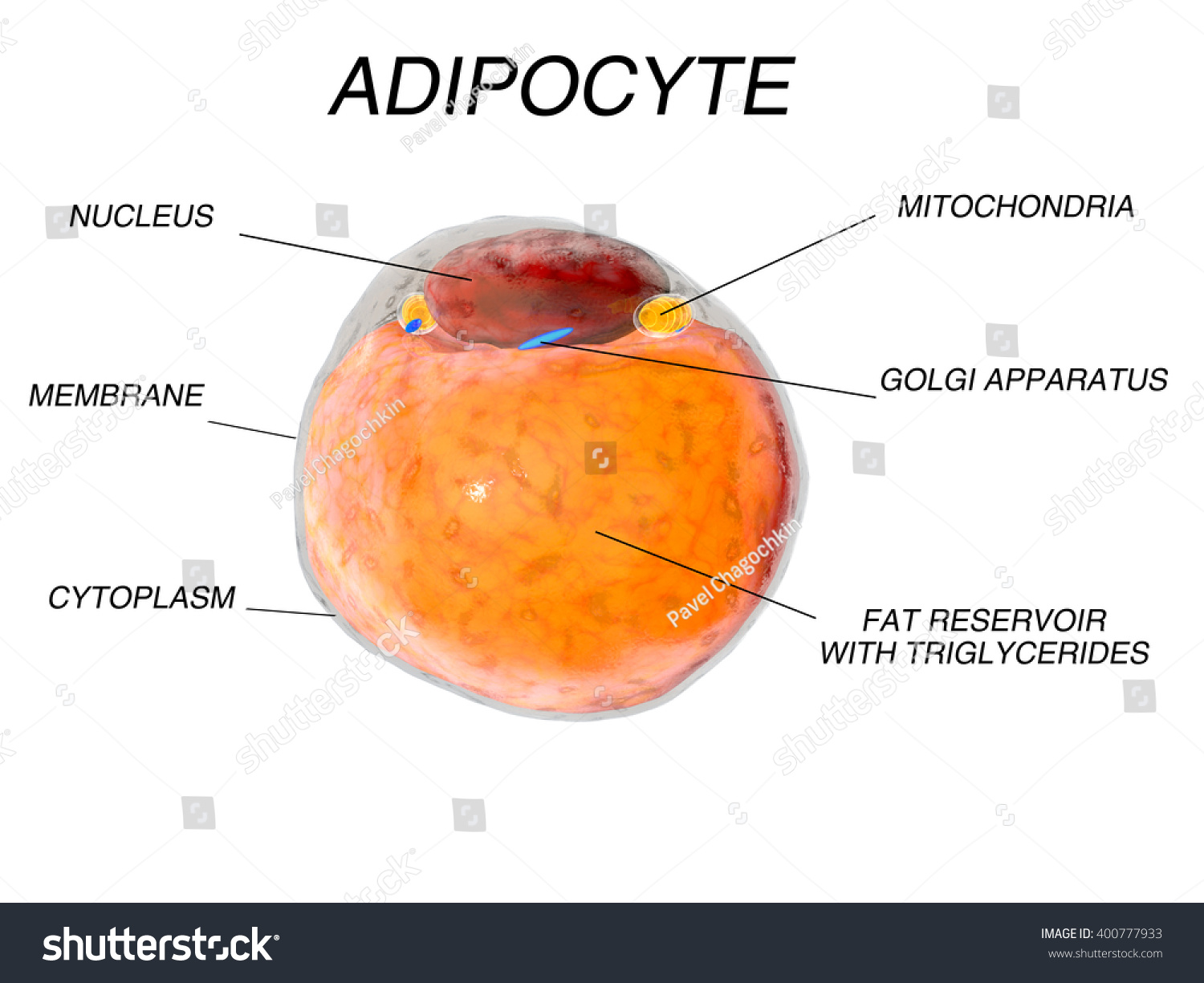
Adipose tissue with labels
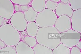
Label-free profiling of white adipose tissue of rats ... We analysed visceral adipose tissue of HCR and LCR using label-free HDMS E profiling. The running capacity of HCR was 9-fold greater than LCR. The running capacity of HCR was 9-fold greater than LCR. Proteome profiling encompassed 448 proteins and detected 30 significant (p <0.05; false discovery rate <10%, calculated using q-values) differences. Adipose connective tissue - Austin Community College District Adipose connective tissue 400X The bar labeled "a" indicates the width of one adipose cell (adipocyte). The light purple dots you see inside the cells are an artifact of process used to make the images, and do not represent real structures. The arrow points to the nucleus of one adipocyte. SC 2115 Anatomy and Physiology I - Middlesex Community College Draw and label Reticular Tissue: reticular fibers form the stroma . E. Adipose Tissue: surrounds heart and kidneys, subcutaneous tissue, and greater omentum. This is the most easily recognized tissue and will be found widely distributed in every organ microscopically studied this year.
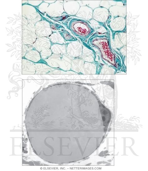
Adipose tissue with labels. Adipose tissue - Vida Private Label Adipose tissue. Is it possible to act on adipose tissue, and how? A treatment aimed at reducing fat deposits and localised oedemas. Fat deposits. Adipose tissue: a fat lot of good? | Society for Endocrinology 3D rendering of a 100-µm thick section of murine adipose tissue in the kidney. Immunofluorescent staining for the lipid droplet protein perilipin is in cyan. A lineage tracing Cre-induced tdTomato fluorescent protein labels a subset of adipocytes red. In the centre, the nucleus of a resident adipose stem cell is labelled green. ©J. Rochford Adipose connective tissue - University of Wisconsin-La ... Adipose connective tissue 1. Cell membrane 2. Cell nucleus 3. Fat vacuoles Adipose connective tissue cells are specialized for fat storage and do not form ground substance or fibers. On prepared slides, adipose tissue appears somewhat like a fish net with white spaces connected together in a network. Adipose Tissue and Loose Connective Tissue: Functions and ... White adipose tissue accounts for about 20-25% of a healthy, non-overweight human's body weight and is used as an energy source. White adipose tissue consists of a single fat droplet.
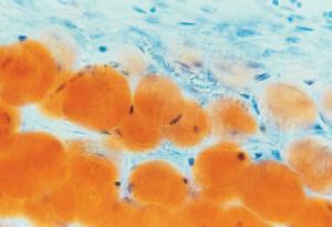
Adipose Tissue Histology - Adipose tissue (labels ... Adipose tissue (labels) - histology slide This histology slide is of adipose tissue. Histology slide courtesy of deltagen.com. Labeled Tissues Flashcards - Quizlet Adipose Tissue Label ground substance, elastic fibers, collagen fibers, fat droplet, specific cells of each tissue Dense ( White) Regular Tissue Label ground substance, elastic fibers, collagen fibers, fat droplet, specific cells of each tissue Hyaline Cartilage Adipose tissue: Definition, location, function | Kenhub Brown adipose tissue labeled (histological slide) In contrast to white adipocytes, brown adipocytes have the appearance of a sponge due to the multiple droplets in the cytoplasm. Groups of adipocytes are divided into lobules by connective septa, which contain a substantial amount of blood vessels and unmyelinated nerve fibers. Off-label use of adipose-derived stem cells - ScienceDirect 1. Introduction. Recent advances in regenerative medicine, particularly the discovery of multipotent, easily accessible stem cells such as adipose-derived stem cells (ASCs), have provided the opportunity to use autologous stem cell transplants as regenerative therapies .Fat is an active and dynamic tissue composed of several different cell types, including adipocytes, fibroblasts, smooth ...
Adipose Tissue - The Definitive Guide| Biology Dictionary Adipose tissue is split into two main types of connective tissue - white and brown - that store and burn energy respectively. White adipose tissue also provide a layer of insulation, while brown adipose is found in too small quantities (in children and adults) to do this. Brown fat does, however, release energy in the form of heat. Adipose Tissue - Composition, Location and Function Adipose, or fat, tissue is loose connective tissue composed of fat cells known as adipocytes. Adipocytes contain lipid droplets of stored triglycerides. These cells swell as they store fat and shrink when the fat is used for energy. Adipose tissue helps to store energy in the form of fat, cushion internal organs, and insulate the body. Adipose Tissue - Meaning, Types, Functions, and FAQs Adipose tissue is a special and different type of connective tissue, mainly composed of fat cells called adipocytes. Adipocytes are classified into three types: white adipocytes, brown adipocytes, and beige adipocytes. Their structure, location, and function are different. Adipose tissue - Wikipedia Adipose tissue, body fat, or simply fat is a loose connective tissue composed mostly of adipocytes. In addition to adipocytes, adipose tissue contains the stromal vascular fraction (SVF) of cells including preadipocytes, fibroblasts, vascular endothelial cells and a variety of immune cells such as adipose tissue macrophages.
How Adipose Tissue Works - Terumo BCT Process Adipose Tissue Into a Purified MSC-Rich Graft in Just 4 Minutes 1. The AdiPrep ® Adipose Concentration System concentrates tissue samples to deliver a graft with high stem cell and nucleated cell counts while significantly reducing excess fluid that contribute to graft volume loss. The resulting purified adipose concentrate can then be ...
Two-photon excited fluorescence of intrinsic fluorophores ... Adipose tissue function has been recognized to exert significant influence on systemic metabolic balance and overall homeostasis health through energy storage via lipid accumulation, direct energy expenditure through substrate oxidation 1,2, and secretion of various signaling and regulatory molecules 3,4.Major health problems such as type 2 diabetes mellitus, cancer, and cardiovascular disease ...
Solved Drag the labels onto the diagram to identify the ... Anatomy and Physiology questions and answers. Drag the labels onto the diagram to identify the parts of the kidney. Reset Help Minor calyx Hilum Ureter Adipose tissue in renal sinus Renal pelvis Renal sinus Major calyx Connection to minor calyx.
Brown-adipose-tissue mitochondria: photoaffinity labelling ... Brown-adipose-tissue mitochondria: photoaffinity labelling of the regulatory site of energy dissipation Abstract Brown-adipose-tissue mitochondria possess an energy-dissipating ion uniport which is inhibited by purine nucleotides.
Solved Label the components of adipose tissue. Fibroblast ... Label the components of adipose tissue. Fibroblast Cell membrane Elastic fiber Nucleus Adipocyte ; Question: Label the components of adipose tissue. Fibroblast Cell membrane Elastic fiber Nucleus Adipocyte
Adipose Tissue Histology Adipose Tissue Histology. Title • File Name • Date • Position. Fat, mouse - histology slide. Fat, mouse - histology slide. Brown adipose tissue (labels) - histology slide. Adipose tissue, mouse - histology slide. Adipose tissue - histology slide. Adipose tissue - histology slide. Adipose tissue - histology slide.
The developmental origins of adipose tissue | Development ... Adipose tissue is formed at stereotypic times and locations in a diverse array of organisms. Once formed, the tissue is dynamic, responding to homeostatic and external cues and capable of a 15-fold expansion. The formation and maintenance of adipose tissue is essential to many biological processes and when perturbed leads to significant diseases.
Skeletal Muscle and Adipose Tissue Responses After Acute ... Skeletal Muscle and Adipose Tissue Responses After Acute Exercise: Comparing Effects of Three Different Intensities (3X) The safety and scientific validity of this study is the responsibility of the study sponsor and investigators.
Photoaffinity labeling of hamster brown adipose tissue ... In this study brown adipose tissue mitochondria were photolabeled with the azido [125I] ACT-CoA derivative with or without inhibitors. SDS gel electrophoresis and autoradiography of the separated proteins revealed exclusive photolabeling of two polypeptides corresponding to the ADP/ATP carrier and uncoupling protein.
Adipose Tissue: What Is It, Location, Function, and More ... Adipose tissue is a specialized type of connective tissue that arises from the differentiation of mesenchymal stem cells into adipocytes during fetal development. Mesenchymal stem cells are pluripotent cells that can transform into various cell types, including fat cells, bone cells, cartilage cells, and muscle cells, among others.
Adipose Tissue: Function, Location & Definition - Video ... Adipose is a loose connective tissue that fills up space between organs and tissues and provides structural and metabolic support. It is part of the nutrient glue that holds us all together....
SC 2115 Anatomy and Physiology I - Middlesex Community College Draw and label Reticular Tissue: reticular fibers form the stroma . E. Adipose Tissue: surrounds heart and kidneys, subcutaneous tissue, and greater omentum. This is the most easily recognized tissue and will be found widely distributed in every organ microscopically studied this year.
Adipose connective tissue - Austin Community College District Adipose connective tissue 400X The bar labeled "a" indicates the width of one adipose cell (adipocyte). The light purple dots you see inside the cells are an artifact of process used to make the images, and do not represent real structures. The arrow points to the nucleus of one adipocyte.
Label-free profiling of white adipose tissue of rats ... We analysed visceral adipose tissue of HCR and LCR using label-free HDMS E profiling. The running capacity of HCR was 9-fold greater than LCR. The running capacity of HCR was 9-fold greater than LCR. Proteome profiling encompassed 448 proteins and detected 30 significant (p <0.05; false discovery rate <10%, calculated using q-values) differences.





Post a Comment for "41 adipose tissue with labels"